Case Report - Bilateral Rehabilitation With Zirconia Implants
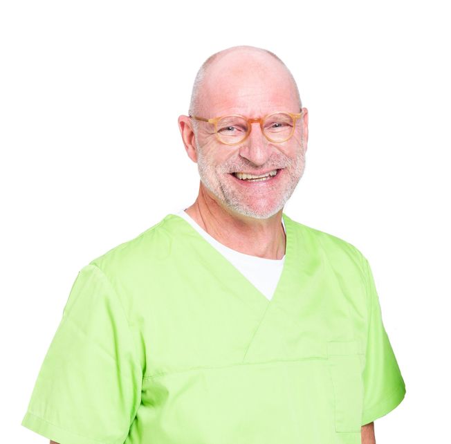
Dr. med. dent. Wolfgang Winges
Curriculum vitae
Since 1988 Dr. Winges has been practicing dentistry in the Hesse region of Bad Hersfeld and is the co-owner of the Winges and Caselitz praxis. He is also the co-inventor of the Patent™ dental implant system. His practice is dedicated to:
• General dentistry
• Implantology & metal-free restorations
• Periodontal therapy
Additionallyheis a lecturerandtrainer for topics encompassing there almofi mplantology and surgery.
Introduction
The 36-year-old patient presented herself for the first time in our practice in May 2019, when she was fitted with removable model cast dentures.
These were rarely worn, as recurrent mucosal inflammation and pressure sores developed repeatedly.
The patient's desire for fixed dentures, also with regard to her age, was very pronounced.
Due to a proven metal incompatibility, a therapy with titanium implants was contraindicated and was expressly rejected by the patient.
Dr. med. dent. Wolfgang Wings
Gerwigstraße 4,36251 Bad Hersfeld www.dr-winges.de
Telephone +43 664 9127454 praxis@dr-winges.de
12 , 2020
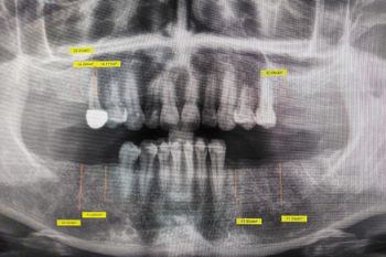
Figure 1:
Preoperative planning by means of OPG scan.
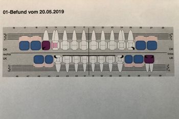
Figure 2:
Tooth status as of May 20, 2019.

Figure 3:
OPG at the time of implant placement.
Therapy
After removal of the extremely pain sensitive teeth in positions 15, 34 and 37, ozonation of the extraction sockets and healing, further planning was done.
A DNA marker germ test was then performed (Hain Lifescience) showing a clear germ load in the green complex.
An adequate antibiotic treatment over 7 days , with 3 of those days applying a 500mg Amoxicillin treatment, improved the oral flora significantly and reduced the bacterial inflammation significantly.
Planning
In addition to study models, an OPG was also used for planning purposes. Due to the extremely good residual bone situation of the non-tooth-bearing alveolar ridges, DVT diagnostics were not necessary.
In our opinion, three-dimensional diagnostics is indicated in cases of greatly reduced and unclear bone supply.
The necessity to determine the vitamin D level before therapy with ceramic implants is controversially discussed.
In our practice we always carry out a chair side measurement of the vitamin D level using the Vitality Health Check.
We aim for a value of 50 ng/ml. For lower values, a substitution with Vit D3 is necessary, which makes sense 3 weeks preop and one week after surgery.
Our patient showed a good vitamin D content of 64 ng/ml.
Implant Therapy
After healing; model and x-ray analysis’ were conducted and planned for the areas of 16, 15, 46, 45, 26 as well as 34 and 36, in order to place ceramic implants (Patent™) of the company Zircon Medical Management (Switzerland).
The Patent™ Dental Implant system has many advantages for such an extensive rehabilitation. The patented manufacturing process ensures a strong and long lasting implant body. Due to this manufacturing process, the surface roughness is unique among ceramic implants designs, is hydrophilic and allows for predictable osseo integration.
In addition, the tissue level design with integrated abutment and a glass fiber post allow for a truly metal free rehabilitation and access to the crown to implant interface for easy home care and hygiene.
After detailed explanation the primary surgery was performed on 27.02.2020 with local anesthesia (Ubistesin 1:100000).
Since the patient has a very broad-based fixed gingiva, it was possible to perform the primary operation minimally invasive, only with mucosal punch.
The advantages of such a procedure are well known.
It is important to choose a punch diameter that is always one size larger than the implant diameter, thus preventing contamination of the rough, patented implant surface of the patented implants by connective tissue.
Of course, the classic procedure with the formation of a mucoperiosteal flap is indicated in cases of reduced bone and mucosa supply.
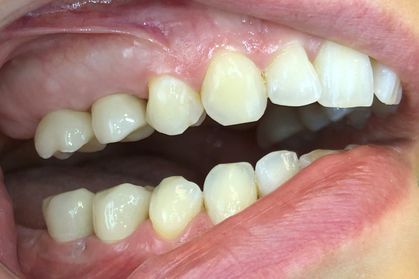
Figure 4:
Restorations in the upper and lower right posterior region.
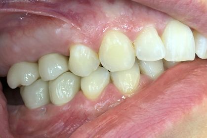
Figure 5:
in occlusion.
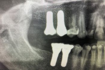
Figure 6:
X-ray control image of the restorations in the right posterior region.
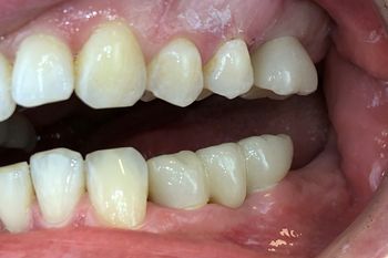
Figure 7:
Restorations in the upper and lower left posterior region.
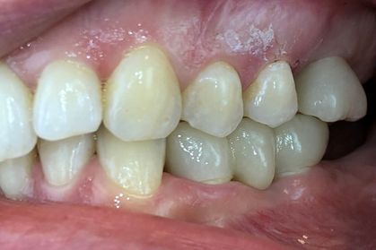
Figure 8:
in occlusion.
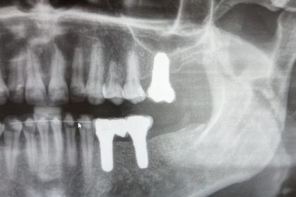
Figure 9:
Radiographic control images of the restorations in the left posterior region.
OP
The following Patent™ implants were inserted:
Standard implant Regio 16,15: Size: 5.0 x 11mm , Regio 36,34,45,46: Size: 4,5 x 11mm , Individual implants Regio 26: Size: 6.0 x 9mm. The insertion torque was 35 Ncm for all implants.
In order to achieve greater primary stability in the maxilla, particularly in bone qualities D3 – D4, we do not use the last drill (underdrilling).
The postoperative radiographic control shows that the implants are seated correctly along the axis.
The patient was instructed to eat only soft food for the next few days. Wound cleaning was done with a very soft wound toothbrush and CHX.
The subsequent wound check on March 5, 2020 showed no abnormalities, the patient was completely free of complaints.
We always perform a Periotest from the Medizintechnik Gulden company (Germany) after 10 weeks to prove successful osseointegration.
Our patient showed negative values on all implants from 1.9 to - 6.9, with slightly lower values in the maxilla. All implants were successfully osseointegrated.
Prothetics
The further prosthetic restoration was carried out on 29.6.2020 by gluing the fiberglass pins into the 3C connection of the patent implants using Rely X Unicem from 3M Espe. This adhesive shows enormously high strength values.
The implants can be restored in the following way:
1. Gluing of the fiberglass pins and preparation according to known rules, followed by impression taking.
2. Direct impression of the 3C connection, grinding of the fiberglass pins in the laboratory and fabrication of the denture. Simultaneous gluing of pin and denture.
3. Scanning of the 3C connection and further procedure in the laboratory.
In the case described above, all fiberglass pins were glued in quadrants and prepared. The impression was taken with Impregum and individualized trays.
The crowns and bridges were fabricated with pritimultidisc ZrO2 High Translucent from the company (pritidenta). This material is characterized by a significantly lower bending strength of > 650MPa and in our opinion is optimally suited for the restoration of ceramic implants.
We pay great attention to contact-free mesial and distal rims and strive for centric contacts.
After color matching and occlusion check the denture could be glued in with Rely X Unicem. The epigingival position of the crown margin makes it much easier to remove excess adhesive. Cementitis" is excluded.
The treatment of the patient was completed with the insertion of a protective splint for the night.
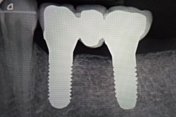
Figure 10:
Radiographic control image of the posterior restorations in the lower left region.
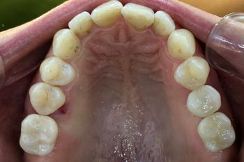
Figure 11:
Occlusal view of the maxillary restorations.
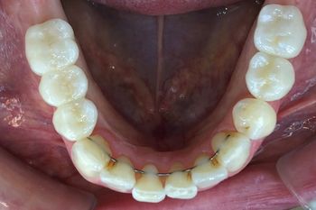
Figure 12:
Occlusal view of the mandibular restorations.
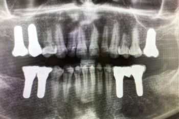
Figure 13:
Final OPG exposure.


